



Meta Description:
If you are considering an MRI for lower back pain in Korea, one of the most common questions is what actually happens before, during, and after the scan. A lumbar spine MRI is designed to examine structures in the lower back, including the lumbar vertebrae, intervertebral discs, spinal canal, nerve roots, and surrounding soft tissues. It can be particularly useful when back pain is associated with persistent leg pain, numbness, weakness, sciatica-type symptoms, or when symptoms do not improve as expected. However, MRI is not automatically necessary for every episode of uncomplicated lower back pain. Clinical evaluation remains important because MRI findings such as disc degeneration or bulging can also appear in people who do not have significant pain.
Hanshin Medipia in Seoul operates a dedicated Radiology Center offering MRI, MRA, MRE, 128-channel MD-CT, and PET-CT examinations. Its imaging center has accumulated extensive experience across multiple diagnostic imaging examinations. Hanshin Medipia also provides English-language information for international patients and operates a health-checkup system that can assist visitors with reservations and examination coordination.
When Is MRI Used for Lower Back Pain?
An MRI for lower back pain is most useful when the goal is to evaluate structures that cannot be seen clearly on a basic X-ray. MRI can provide detailed images of the lumbar discs, nerve roots, spinal canal, vertebral structures, ligaments, and soft tissues around the spine.
For uncomplicated lower back pain that has only been present for a short period and does not include neurological warning signs, immediate MRI is often not necessary. Many cases improve with conservative management.
MRI becomes more relevant when symptoms persist, worsen, or suggest possible nerve or spinal canal involvement. Lumbar MRI is also commonly considered when treatment planning depends on identifying the location of nerve compression or other structural changes.
• Persistent lower back pain
• Pain radiating from the lower back into the buttock or leg
• Sciatica-type symptoms
• Leg or foot numbness
• Tingling in the lower extremities
• Progressive weakness
• Suspected lumbar disc herniation
• Suspected spinal stenosis
• Symptoms that persist despite treatment
• Evaluation before certain procedures or surgery
What Does a Lumbar Spine MRI Check?
A lumbar spine MRI usually focuses on the lower section of the spine, including the lumbar vertebrae from L1 through L5 and the lumbosacral junction around L5-S1.
The examination can show the condition of the intervertebral discs, spinal canal, nerve roots, vertebral bone marrow, ligaments, and surrounding soft tissues.
This makes lumbar MRI particularly useful when lower back pain is accompanied by symptoms traveling into the leg.
Common areas evaluated include:
• L1-L2
• L2-L3
• L3-L4
• L4-L5
• L5-S1
• Lumbar spinal canal
• Nerve roots
• Intervertebral discs
• Neural foramina
• Surrounding soft tissues
The lower lumbar levels, particularly L4-L5 and L5-S1, are frequently evaluated because disc degeneration and nerve-root compression commonly occur in these areas.
MRI for Back Pain: What to Expect Before the Scan
If you are searching for MRI for back pain what to expect, preparation is usually straightforward.
Before entering the MRI room, the medical team needs to know whether you have any implanted medical devices, previous surgery, metal in your body, or other conditions that could affect MRI safety.
Because MRI uses a very strong magnetic field, metal objects must generally be removed before the examination.
You should tell the medical team if you have:
• A pacemaker or implanted cardiac device
• Cochlear implants
• Aneurysm clips
• Artificial joints or metal plates
• Surgical screws or other implanted metal
• Vascular stents
• Previous surgery
• Metal fragments in the body
• Kidney disease
• Pregnancy or possible pregnancy
• Claustrophobia
• Previous reactions to MRI contrast material
Jewelry, watches, removable metal accessories, hearing aids, and other objects that may be affected by the magnetic field should also be removed before the examination.
What Happens During a Lumbar MRI?
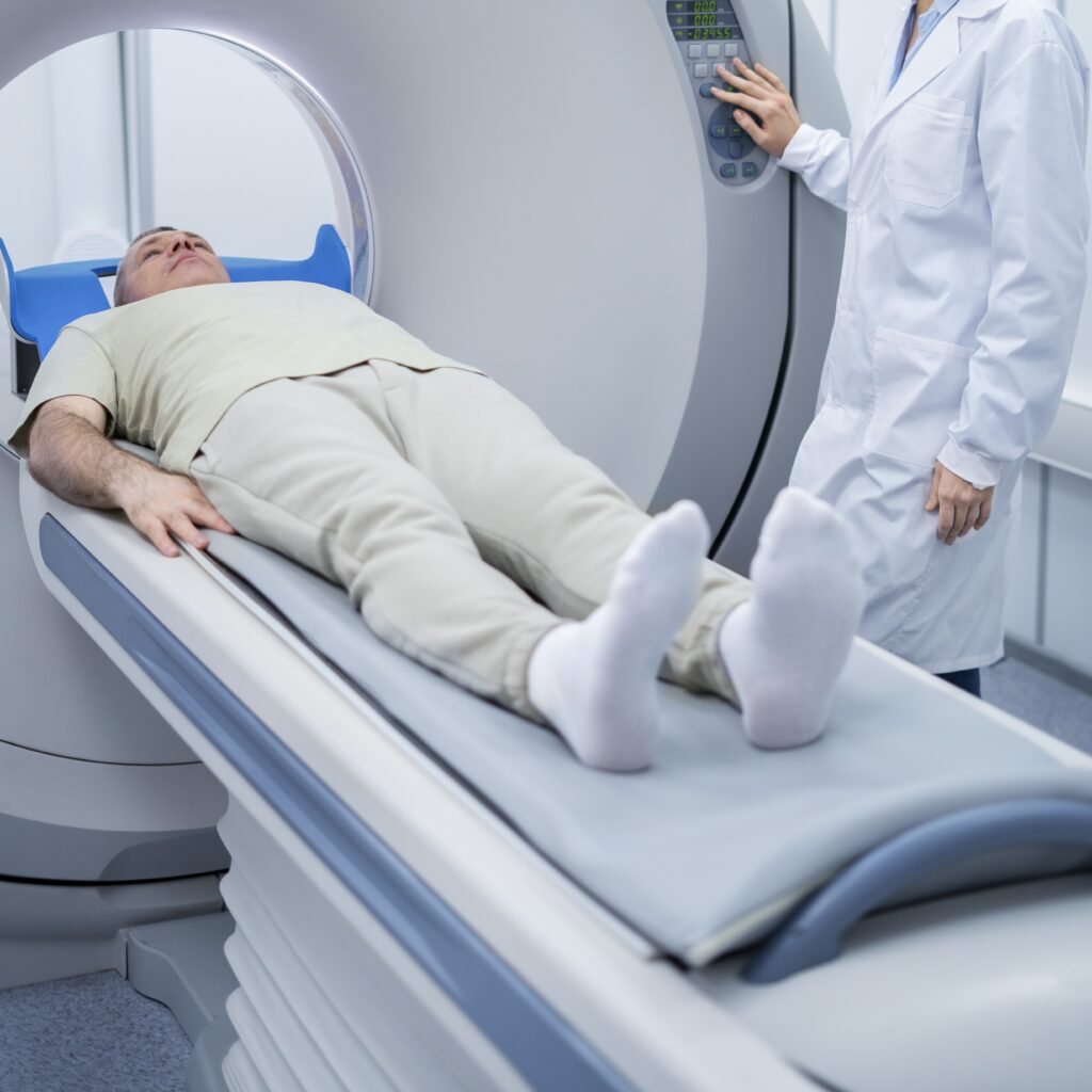
During the examination, you lie on a flat MRI table that moves into the scanner.
For a lower back MRI, you will usually lie on your back while the lumbar region is positioned for imaging.
The most important part is remaining still. Movement can reduce image quality and may require portions of the examination to be repeated.
MRI scanners also make loud tapping, knocking, or humming sounds while images are being acquired. Ear protection such as earplugs or headphones is typically used.
During an MRI:
• You lie flat on the examination table
• The lumbar area is positioned for imaging
• The table moves into the MRI scanner
• You need to remain as still as possible
• The scanner makes repeated loud sounds
• Earplugs or hearing protection may be provided
• Staff monitor the examination from another room
• Communication with the technologist remains possible
A lumbar MRI commonly takes around 30 to 60 minutes, although the exact time can vary according to the examination protocol and whether additional sequences or contrast are required.
Does an MRI for Lower Back Pain Hurt?
The MRI itself is not considered painful.
For patients who already have significant lower back pain, the most uncomfortable part may be lying flat and remaining still for the duration of the scan.
If staying in one position is difficult because of pain, it is helpful to tell the staff before the scan begins.
Some patients also experience discomfort because of the enclosed space inside the scanner. If you have significant claustrophobia, this should be discussed before your appointment.
• MRI itself does not involve surgical pain
• Remaining still may be uncomfortable with severe back pain
• The scanner can feel enclosed
• Loud noises are normal during scanning
• Tell staff beforehand if you have claustrophobia
• Communication with staff is available during the examination
Does Lumbar Spine MRI Use Radiation?
MRI does not use ionizing radiation.
Instead, magnetic resonance imaging uses a strong magnetic field, radiofrequency energy, and computer processing to create detailed images.
This is different from X-ray and CT examinations, which use ionizing radiation.
For patients who are comparing imaging options in Korea, the distinction is useful:
• MRI → no ionizing radiation
• MRA → magnetic resonance-based blood vessel imaging
• CT → uses X-rays
• MD-CT → uses X-rays with multidetector CT technology
• PET-CT → includes CT and nuclear medicine imaging
Hanshin Medipia operates MRI as well as 128-channel MD-CT and PET-CT, allowing different imaging methods to be selected according to the medical question being evaluated.
Will Contrast Be Needed for a Back MRI?
Many routine lumbar spine MRI examinations are performed without contrast.
Contrast-enhanced MRI may be used when the medical team needs additional information, such as in selected cases involving previous spine surgery, suspected infection, inflammation, or certain tumors.
If contrast is being considered, medical history and kidney function may be relevant.
The decision should be based on the reason for the MRI rather than assuming that contrast is always necessary.
• Many routine lumbar MRI examinations do not require contrast
• Contrast may be used for selected medical questions
• Previous spine surgery can affect the protocol
• Kidney problems should be disclosed beforehand
• The radiology team determines whether contrast is appropriate
What Can a Lower Back MRI Find?
A lumbar MRI can identify structural changes in and around the lower spine.
Possible findings may include:
• Disc bulging
• Disc protrusion or herniation
• Lumbar spinal stenosis
• Nerve-root compression
• Degenerative disc changes
• Facet-joint degeneration
• Vertebral abnormalities
• Inflammation
• Infection
• Certain tumors
• Postoperative changes
However, an important point for anyone researching an MRI for back pain is that not every MRI abnormality explains the patient’s symptoms.
Age-related disc changes and degeneration can appear on MRI even in people without pain. This is why an MRI result should be interpreted together with symptoms, physical examination findings, and neurological findings rather than in isolation.
When Should Back Pain Be Evaluated More Urgently?
Some lower back symptoms require more urgent medical evaluation rather than routine screening.
Seek prompt medical assessment when lower back pain is associated with symptoms such as:
• New loss of bladder control
• New loss of bowel control
• Numbness around the groin or saddle area
• Rapidly worsening leg weakness
• Significant difficulty walking
• Severe back pain after major trauma
• Back pain accompanied by fever
• Back pain with a history of cancer
• Progressive neurological symptoms
These symptoms can raise concern for conditions that may require urgent imaging or treatment.
Hanshin Medipia Lumbar Spine MRI in Korea
Hanshin Medipia offers lumbar spine MRI as one of its MRI examinations.
The center also provides musculoskeletal MRI examinations that can include cervical or lumbar spine MRI along with selected joint MRI examinations.
For someone experiencing persistent lower back symptoms, lumbar spine MRI can help evaluate discs, spinal structures, and nerve-related anatomy.
Hanshin Medipia’s Radiology Center is equipped for several advanced imaging examinations, including MRI, MRA, MRE, MD-CT, and PET-CT.
• Lumbar spine MRI available
• Cervical spine MRI available
• MRI integrated with broader health screening
• Dedicated Radiology Center
• MRI, MRA, MRE, MD-CT, and PET-CT available
• International patient support available
Other Hanshin Medipia MRI Tests
Hanshin Medipia MRI options extend beyond lower back imaging.
Depending on the patient’s health concerns or screening goals, the center can provide the following examinations:
• Brain MRI
• Cerebrovascular MRA
• Cervical spine MRI
• Lumbar spine MRI
The center also offers special MRI examinations in the afternoon.
These include:
• Hip joint MRI
• Wrist MRI
• Left or right shoulder MRI
• Left or right ankle MRI
• Left or right knee MRI
Hanshin Medipia also offers:
• Pancreas MRI — afternoons only
• Whole-body MRI — afternoons only
Hanshin Medipia Whole-Body MRI
Hanshin Medipia also offers a Whole-body MRI option in the afternoon.
This specific program takes approximately 35 minutes and combines:
• Brain MRI
• Brain MRA
• Cervical spine MRI
• Lumbar spine MRI
For international patients who want to examine both the cervical and lumbar spine while also undergoing brain and cerebrovascular magnetic-resonance imaging, this can make it possible to coordinate several MRI regions within one program.
It is important to understand the scope of the program correctly.
Hanshin Medipia’s Whole-body MRI refers to this specific combination of brain MRI, brain MRA, cervical spine MRI, and lumbar spine MRI. It should not automatically be interpreted as an MRI examination of every organ in the body.
Brain MRI and Cerebrovascular MRA
Hanshin Medipia also provides brain MRI and cerebrovascular MRA.
Although the technologies are related, MRI and MRA serve different purposes.
Brain MRI focuses primarily on brain structures and tissue, while cerebrovascular MRA is designed to visualize blood vessels within and around the brain.
Hanshin Medipia offers brain MRI and brain MRA among its neurological screening options.
• Brain MRI → evaluates brain structures
• Brain MRA → evaluates cerebral blood vessels
• Both can be coordinated in selected screening programs
• Whole-body MRI also includes brain MRI and brain MRA
Cervical Spine MRI
Not all spinal symptoms originate in the lower back.
A cervical spine MRI focuses on the neck and the upper spinal cord.
It may be considered when symptoms involve the neck, shoulders, arms, hands, or cervical nerve structures.
Typical symptoms that can lead to cervical spine evaluation include:
• Persistent neck pain
• Shoulder or arm pain
• Hand numbness
• Arm tingling
• Upper-limb weakness
• Suspected cervical disc disease
• Suspected cervical spinal stenosis
Hanshin Medipia offers both cervical spine MRI and lumbar spine MRI, allowing the appropriate spinal region to be selected according to symptoms.
Special MRI for Joint Problems
Back pain is not always caused by the lumbar spine alone.
Hip problems, for example, can sometimes produce pain around the lower back, buttock, or groin area.
For this reason, the location and pattern of symptoms are important when selecting an imaging examination.
Hanshin Medipia’s afternoon special MRI options include:
• Hip joint
• Wrist
• Left or right shoulder
• Left or right ankle
• Left or right knee
These targeted examinations allow a specific joint to be evaluated when the primary problem is not suspected to originate from the lumbar spine.
Pancreas MRI
Hanshin Medipia also offers pancreas MRI in the afternoon.
This examination has a very different purpose from a lumbar spine MRI and is intended for pancreatic and related abdominal evaluation.
The availability of several MRI examinations within one health-screening center can be practical for international visitors who want to coordinate different diagnostic or screening needs during their time in Korea.
• Pancreas MRI available
• Afternoon scheduling
• Separate purpose from spine MRI
• Can be coordinated with broader health screening when appropriate
Why International Patients Consider Hanshin Medipia
One of the main strengths of Hanshin Medipia is the ability to combine imaging examinations with a broader health-screening system.
The center operates specialized areas for radiology, ultrasound, laboratory medicine, endoscopy, women’s health, and thyroid screening.
Its Radiology Center includes MRI, MRA, MRE, 128-channel MD-CT, and PET-CT.
Hanshin Medipia also provides English-language information and support for international patients, including assistance with reservations and examination coordination.
• Dedicated Radiology Center
• MRI, MRA, MRE, MD-CT, and PET-CT
• Comprehensive health-screening system
• International patient support
• English-language information available
• Multiple examinations can be coordinated according to the schedule
What International Patients Should Prepare
International visitors planning an MRI for lower back pain in Korea should prepare medical and travel information before the appointment.
It can be helpful to provide:
• Main back-pain symptoms
• Exact location of pain
• Whether pain radiates into the buttock or leg
• Numbness or weakness
• Duration of symptoms
• Previous MRI or CT examinations
• Previous spine surgery
• Implanted medical devices
• Metal implants
• Current medications
• Relevant medical conditions
• Arrival and departure dates in Korea
If your time in Korea is limited, providing your travel dates in advance can also help with scheduling.
What to Expect After the MRI
After a routine lumbar MRI without sedation, there is generally no special recovery period.
Patients can usually return to normal activities unless they have been given separate medical instructions.
The MRI images then need to be interpreted by a radiology specialist.
The result should be considered together with your symptoms rather than simply focusing on whether the report contains terms such as “degeneration,” “bulging,” or “disc protrusion.”
In other words, the most useful question is not simply, “Is something abnormal on MRI?” but rather, “Does this MRI finding match my symptoms?”
• Routine MRI usually requires no recovery period
• Images are interpreted after the examination
• Findings should be correlated with symptoms
• Degenerative findings do not always explain pain
• Follow-up consultation may be required
MRI for Lower Back Pain in Korea: What to Expect Overall
For patients searching MRI for back pain what to expect in Korea, the overall process is relatively straightforward.
You first confirm whether lumbar spine MRI is the appropriate examination for your symptoms. Before the scan, MRI safety information is reviewed and metal objects are removed.
During the scan, you lie still while the MRI machine acquires detailed images of the lower spine. You will hear loud noises, but communication with the medical staff remains possible.
Afterward, the images are reviewed and interpreted in the context of your symptoms.
Hanshin Medipia offers lumbar spine MRI as well as cervical spine MRI, brain MRI, cerebrovascular MRA, special joint MRI, pancreas MRI, and a Whole-body MRI program combining brain MRI, brain MRA, cervical spine MRI, and lumbar spine MRI.
For international patients who want to combine an MRI with other health-screening examinations in Korea, having several imaging and screening services within one center can make coordination easier.
Hanshin Medipia and Kosmedi
Hanshin Medipia is an official Kosmedi(Soon to be Kormpath) partner clinic. International patients who want to learn more about Hanshin Medipia MRI, lumbar spine MRI, cervical spine MRI, brain MRI, MRA, Whole-body MRI, or other health-screening examinations can contact Kosmedi for assistance. Kosmedi can help confirm examination information, available appointment dates, preparation details, and arrange an actual appointment with Hanshin Medipia.
• WhatsApp: +82 10 9948 7172 | https://wa.me/821099487172
• Email: Kosmedisns@gmail.com
• LINE ID: kosmedi
• Instagram: @kosmedi.official | https://www.instagram.com/kosmedi.official/
• X: @KosmediOfficial | https://x.com/KosmediOfficial

